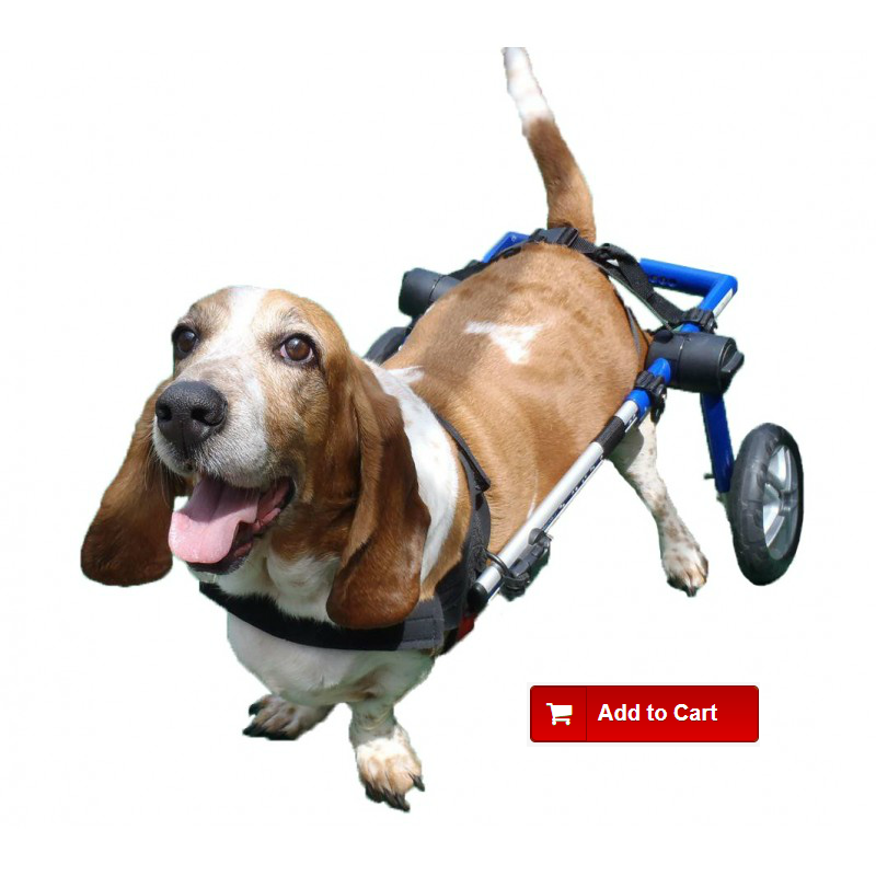
A herniated disc is a degenerative disease of the intervertebral discs and is one of the most common diseases that dogs suffer from when showing difficulty moving the hind legs. Compression of the spinal cord occurs when the disc material leaves the spinal canal (extrusion) or bulges (protrusion). This phenomenon usually causes pain and dysfunction in the spinal cord that can be reflected on different levels; lack of coordination in movement, the dog stops walking, crawling or make costly moves (paresis or paralysis of the limbs), and problems for urination and defecation. To combat the pain, the animal adopts antalgic postures like keeping the head down or arching the dorsal (back) in the area of injury.
This condition can manifest at any intervertebral disc but often this type of injury is found in segments of the cervical spinal and thoracolumbar.
Classification of herniated discs (according to hansen)
Type I Hansen are those corresponding to chondrodystrophoid breeds (small, long spine and short‑legged) such as the poodle, dachshund, Pekingese, cocker, etc., in young animals 2-6 years of age. Chondroid degeneration of the nucleus pulposus occurs with a possible calcification (chondroid metaplasia). The core turns into cartilaginous material, hardens and causes the rupture of the disc’s dorsal fibers and the material to leave the spinal canal (extrusion into the spinal canal) providing a sharp focal compression. It is caused by abrupt movements in the spine such as those resulting from jumps, falls, bumps or climbing on and off the sofa. The compression is acute although the problem may be due to an acute cause or the evolution of micro-traumas.
Type II Hasen disc degeneration corresponds to large breeds that are nonchondrodystrophoid such as Boxers, Labradors, German Shepherds, Rottweilers, etc., in adult animals from 5 to 12 years old. Evolution of this type is slow throughout the dog’s life and problems appear later on. A gradual protrusion of the annulus fibrosus of the intervertebral disc content that has degenerated over time (fibrous metaplasia) is generated. The material is intact; a focal, slow and progressive compression occurs (myelopathy).
It is possible for noncondrodystrophoid breeds to contract Hansen type I disc degeneration at any age.
Type III Hansen disc degeneration refers to an acute, severe extrusion followed by progressive myelomalacia that in many cases leads to the death of the animal.
Clinical Symptoms Displayed
- Pain, the animal adopts antalgic postures due to inflammatory responses (lowers the head and curves the back).
- Decreased proprioception, the animal has a "forgotten" paw and is unable to properly place the pad in contact with the ground, lack of coordinated movements, harsh movements or crawling (paresis or paralysis), difficulty maintaining balance.
- A loss of sensitivity occurs in the injured area and extremities.
- Problems with urinary and/or fecal incontinence or retentions.
- Within a few days an alteration of the muscle tone occurs along with decrease in muscle mass and strength.
To diagnosis herniated discs, the veterinarian must be informed of the dog's history, breed, age, any clinical symptoms the animal presents and they will also perform a neurological examination.
An X-ray of the spine will show whether there is decreased intervertebral space, however it is impossible to be sure of the condition of intervertebral discs unless they are calcified, therefore, it is difficult to see the herniated material. In such cases, a myelogram is generally performed.
Myelography is a technique that introduces an iodized contrast around the spine (subarachnoid area) in order to see its border. It is possible to see where there is compression by examining the myelography.
There are other complementary methods such as CT (Computed Tomography) and Nuclear Magnetic Resonance with which you can also diagnose the exact point where the hernia has occurred.
What to do if Your Dog Has a Herniated Disc


